
About this procedure
Two-Jaw Surgery is an advanced corrective procedure that repositions both the upper jaw (maxilla) and lower jaw (mandible) to improve facial balance, bite alignment, and overall function.
It is often recommended for patients with significant jaw asymmetry, protrusion, retrusion, facial imbalance, or bite problems that cannot be corrected with orthodontic treatment alone.
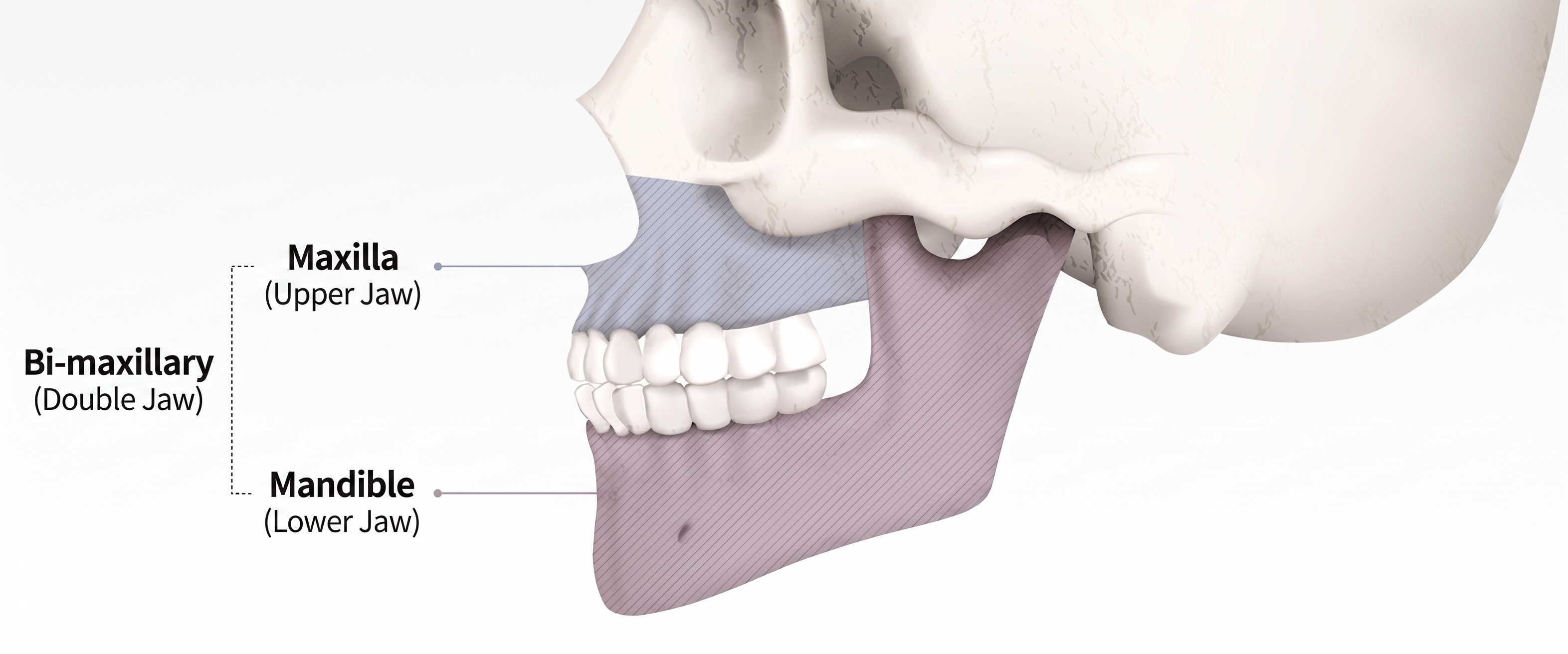
Malocclusion / Prognathism (Lantern Jaw)
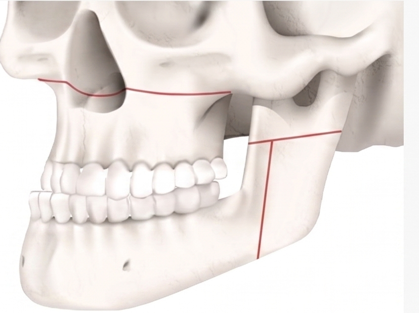
The upper (maxilla) and lower (mandible) jaws are cut.
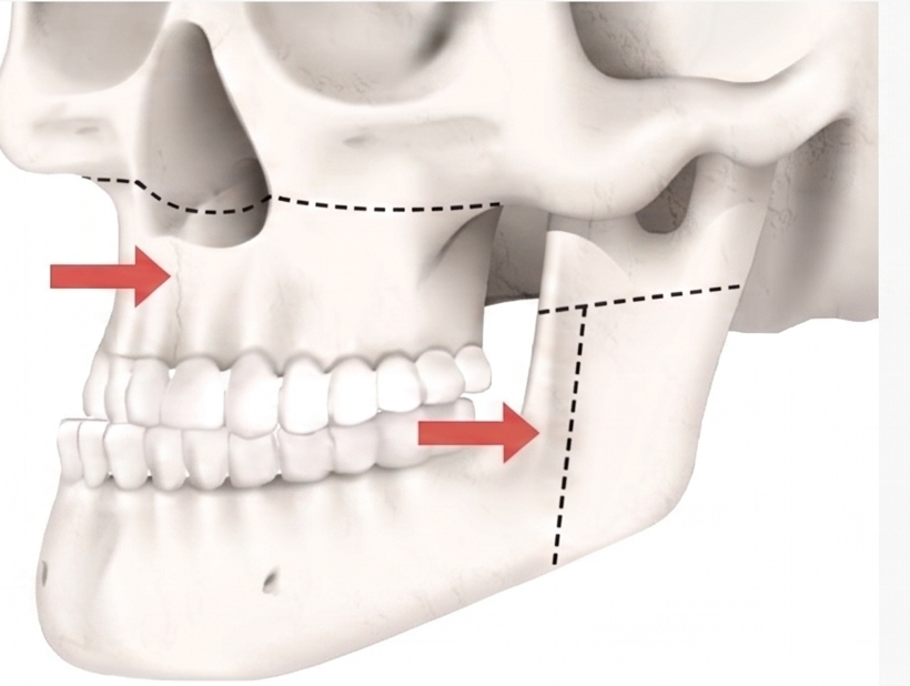
The cut upper and lower jaw bones are moved according to the surgical plan to correct the bite.
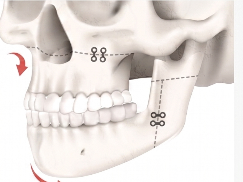
The cut bones are firmly fixed in place so they do not move, completing a balanced face with a correct bite.
Facial Asymmetry
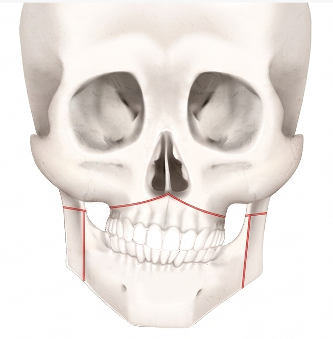
The upper (maxilla) and lower (mandible) jaws are cut (osteotomized).
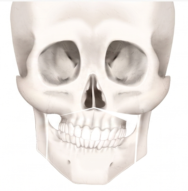
The cut upper and lower jaw bones are moved according to the surgical plan to correct the bite.
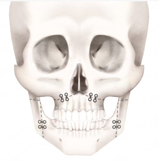
The cut bones are firmly fixed to prevent movement, completing a balanced face with a correct bite.
Two-Jaw Surgery (Bimaxillary Orthognathic Surgery) — Procedure
1. Initial Consultation & Diagnosis
Surgeon and orthodontist evaluate bite alignment, facial symmetry, and the skeletal relationship between the upper jaw (maxilla) and lower jaw (mandible).
3D CT scan, panoramic X-ray, and digital bite analysis are used to map exact bone positions and plan movements.
Photos and facial measurements are taken to plan the aesthetic outcome alongside the functional correction.
2. Pre-Surgical Orthodontics
Braces are placed 6–18 months before surgery to align the teeth within each jaw individually.
This step does not fix the bite yet — it prepares the teeth so that once the jaws are repositioned surgically, they fit together properly.
Surgeon and orthodontist coordinate closely throughout this phase, with regular check-ins to track progress.
3. Surgical Planning
Using imaging data, the surgical team creates a precise plan (often with 3D-printed surgical guides / splints) mapping exactly how many millimeters and in which direction each jaw will move.
A custom “bite splint” is made to guide the jaws into the correct final position during surgery.
4. Anesthesia
Performed under general anesthesia, typically in a hospital operating room.
5. Upper Jaw (Maxilla) Surgery — Le Fort I Osteotomy
An incision is made inside the upper lip / gum line (no external scar).
The upper jawbone is cut horizontally above the teeth roots, fully separating it from the skull.
The maxilla is then repositioned (moved forward, back, up, down, or rotated) according to the surgical plan.
It is secured in its new position using titanium plates and screws.
6. Lower Jaw (Mandible) Surgery — Bilateral Sagittal Split Osteotomy (BSSO)
Incisions are made inside the mouth near the back molars on both sides.
The mandible is carefully split along a natural plane on each side, separating the front tooth-bearing portion from the back jaw-joint portion.
This allows the front part of the jaw (with teeth) to be moved forward, backward, or rotated independently of the jaw joints.
It is also secured with titanium plates / screws.
7. Bite Verification
The custom surgical splint is used intraoperatively to confirm the teeth fit together correctly in the new position.
The surgeon checks bite alignment and facial symmetry before finalizing bone fixation.
8. Possible Additional Procedures (Same Surgery)
Genioplasty (chin repositioning) is often done simultaneously if the chin also needs adjustment to match the new jaw position.
Soft-tissue adjustments around the lips / nose base may be made if jaw movement affects the facial profile.
9. Closing
Incisions inside the mouth are closed with dissolvable sutures.
No external incisions, so there is no visible scarring.
Benefits
Functional Benefits (Primary Focus)
Corrects significant bite misalignment (malocclusion) — overbite, underbite, crossbite, or open bite
Repositions both the upper jaw (maxilla) and lower jaw (mandible) simultaneously for proper occlusion
Improves chewing function and reduces uneven wear on teeth from bite misalignment
Can significantly improve obstructive sleep apnea by advancing the jaws and increasing airway space
Reduces TMJ (jaw-joint) strain caused by chronic bite misalignment
Improves speech clarity in cases where severe jaw discrepancy was affecting articulation
Corrects facial asymmetry caused by skeletal jaw discrepancy (not just soft tissue)
Aesthetic Benefits
Dramatically improves facial profile and balance by correcting the underlying bone structure, not just surface appearance
Can correct a recessed chin / midface (Class II) or a protruding lower jaw (Class III “underbite” look)
Often improves lip seal and resting mouth position if jaw discrepancy was causing an open bite or strained lip closure
Creates more harmonious facial thirds (upper, mid, lower-face proportion)
Frequently combined with genioplasty or V-line contouring for a fully refined result